Lenoir’s spur, a mischievous manifestation of pain under the heel, is an unwelcome companion for many patients entering a podiatric clinic. But what exactly is Lenoir’s spur, this source of discomfort that can so greatly affect quality of life? A journey into the history and anatomy of the foot is necessary to demystify this ailment.
Historically, Lenoir’s spur is a term dating back to the 20th century, referring to a bony outgrowth that forms on the calcaneum, the heel bone. Anatomically, this pathology is related to inflammation of the plantar fascia, the robust band of tissue that supports the arch of the foot, whose health is crucial for the biomechanics of walking and running. Often associated with plantar fasciitis, Lenoir’s spur is closely linked to the individual’s foot type, such as flat feet or those with a high plantar arch.
In podiatric consultation, the evaluation of Lenoir’s spur begins with a thorough anamnesis and meticulous physical examination. Symptoms experienced, such as heel pain and plantar fascia inflammation, guide the podiatrist in their diagnosis. The patient often describes pain particularly felt during the first steps after rest or a prolonged period of physical activity.
Symptoms of Lenoir’s Spur
The symptoms of Lenoir’s spur, or calcaneal spur, go beyond simple plantar pain. Typically, the pain felt is often described as a nail-stabbing sensation in the heel on the first step in the morning or after a period of rest. This pain may subside after a few steps but tends to intensify over the course of the day and after prolonged periods of standing, walking, or stress on the heel. Numbness may occasionally accompany the pain, while night cramps may also signal excessive tension of the plantar fascia related to Lenoir’s spur.
Causes
The causes are multiple, athletes, for example, may develop Lenoir’s spur following repeated movements causing overload on the plantar fascia. Capsulitis, inflammation of the capsules surrounding the joints, is not uncommon in this active population. Moreover, aging leads to changes in the tissues of the foot, increasing the risk in older people.
The biomechanics of the foot is a key factor in the development of Lenoir’s spur. Atypical walking, excessive pronation where the foot rolls inward, or supination where it rolls outward, can create unusual tension on the plantar fascia. This can, in turn, lead to inflammation and the formation of a bony outgrowth on the heel. Foot misalignment can also influence the distribution of pressure on joints and soft tissues, increasing the risk of plantar fasciitis and, consequently, Lenoir’s spur.
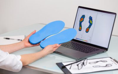
Plantar orthotics are designed to correct biomechanical imbalances of the foot and redistribute body weight more evenly. They are often customized to fit the unique shape of the patient’s foot, providing support to the plantar arch and relief from tension on the plantar fascia. Specialized shoes, meanwhile, may include features like shock-absorbing soles, supportive arches, and materials that adapt to the shape of the foot, thus minimizing the risks of pressure that can lead to Lenoir’s spur.
Genetics also plays a role, as does the choice of footwear – often, poorly fitting shoes can be a source of problems. And let’s not overlook the impact of a demanding job requiring prolonged standing or walking, which can contribute to the onset of this pathology.
Radiology
Medical technology has revolutionized the field of podiatry, particularly in the diagnosis and treatment of Lenoir’s spur. Radiography, ultrasound, and magnetic resonance imaging (MRI) are the cornerstones of diagnostic imaging in the evaluation of this condition.
- Radiography: Radiography is one of the first imaging methods used to visualize Lenoir’s spur. X-rays can detect bony outgrowths on the calcaneum – radiological evidence of Lenoir’s spur. However, it is important to note that the presence of a bony spur in radiography does not always correlate with the patient’s pain symptoms. Some patients may have a significant spur without pain, while others may suffer severe pain with only minimal changes visible on X-ray.
- Ultrasound: This imaging modality uses sound waves to produce images of the internal structures of the foot. Ultrasound is particularly useful for assessing the condition of the plantar fascia itself, revealing its thickness, integrity, and inflammation. It can also help differentiate Lenoir’s spur from other sources of heel pain such as cysts or ruptures of the fascia.
- MRI: For a more detailed evaluation, MRI can be used. It is particularly useful for visualizing both bony structures and soft tissues with a high level of detail. This can be crucial in cases where symptoms persist despite conservative treatments, or when there are doubts about the presence of other pathologies, such as soft tissue lesions, which are not always visible in radiography or ultrasound.
Prevention
In terms of prevention, the use of plantar orthotics can be beneficial, especially for flat feet or poorly defined plantar arches. These devices can evenly distribute weight across the plantar arch, thereby reducing tension on the plantar fascia.
To prevent plantar fasciitis and, by extension, Lenoir’s spur, several strategies can be adopted. At-risk individuals, such as athletes, can benefit from specific stretching exercises for the Achilles tendon and plantar fascia, while older people should focus on maintaining flexibility and strength in the foot. Shoes play a major role: well-fitted shoes with adequate arch support and heel cushioning can decrease tension on the plantar fascia.
Treatment
Home treatment often includes stretching exercises for the Achilles tendon and plantar fascia, as well as applying ice to reduce inflammation. Relief of Lenoir’s spur can also be facilitated by wearing adequately supportive shoes and modifying physical activities to reduce pressure on the heel.
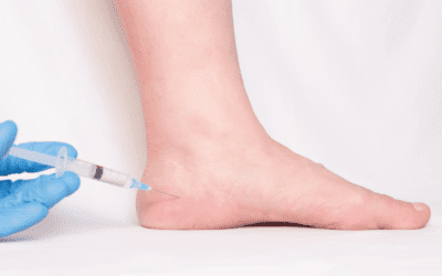
Treatment options for Lenoir’s spur are varied. Traditional treatments include rest, ice, stretches, non-steroidal anti-inflammatory drugs (NSAIDs), and corticosteroid injections. In comparison, shockwave therapy is a non-invasive method that uses sound waves to promote tissue healing. It has been lauded for its effectiveness in reducing pain and improving function without the risks associated with more invasive procedures. For stubborn cases, surgery may be considered, but it is generally seen as a last option due to associated risks and recovery time.
Another expanding technological area is that of shoes and orthotics manufactured by 3D printing techniques, offering unprecedented customization to fit the exact contours of the patient’s foot, thus providing optimal support and comfort, which can prevent and treat Lenoir’s spur by reducing pressure exerted on the heel and plantar fascia.
The podiatrist has a range of treatments for Lenoir’s spur at their disposal. After making their evaluation, they may propose custom orthotics, shockwave therapy to promote tissue healing, or corticosteroid injections to control inflammation. In some cases, surgical intervention may be considered to remove the bony outgrowth.
Beyond diagnosis, podiatric technologies also extend to treatment, with the adoption of advanced techniques such as shockwave therapy. This non-invasive technology uses acoustic pulses to stimulate tissue healing in the affected area. Shockwaves can induce tissue regeneration and neovascularization, which can reduce pain and promote recovery of the plantar fascia.
Podiatric treatments are essential for managing the symptoms of Lenoir’s spur, but prevention remains the best approach. Wise shoe choices, weight management, and attention to early signs of plantar fasciitis can greatly contribute to foot health.
In conclusion, although Lenoir’s spur is a common condition
Proper management by a podiatrist can greatly improve the daily lives of patients. Prevention through good practices and prompt intervention in case of heel pain or other symptoms are key to keeping this pathology at bay and maintaining the health of our feet, these foundations that carry us.


